Foot Muscles Mri Anatomy / Mri Ankle Anatomy Ankle Anatomy Foot Anatomy Anatomy : 3 articles feature images from this case.. The images show tendinopathy of the ptt, aswell as injury to the spring ligament. Here, you will find an overview of the different structures that make up the various aspects of foot anatomy, how they fit together and what can go. Foot and ankle anatomy is quite complex. Learn anatomy faster and remember everything you learn. Radiologists perform ankle imaging to assess injuries of the foot and ankle anatomy.
Cross section of the foot with anatomical structures labeled as arteries, muscles (dorsal interossei, plantar interossei, lombrical, extensor digitorum anatomy of the whole human body : Ayak ve ayak bileği kesitsel mri anatomisi ve hastalıkları. There are around 650 skeletal muscles within the typical human body. Involved early gray = muscle: The feet are flexible structures of bones, joints, muscles, and soft tissues that let us stand upright and perform activities like walking, running, and jumping.
17 Shoulder Anatomy Ideas Shoulder Anatomy Anatomy Mri from i.pinimg.com Almost every movement in the body is the outcome of muscle contraction. The medial muscles of the foot sole have various tasks: In magnetic resonance imaging (mri) of the elbow, patients are imaged in the supine position or in the prone position with the arm overhead. Also explore over 8 similar quizzes in this category. Choose from 500 different sets of flashcards about human anatomy muscles on quizlet. The muscles acting on the foot can be divided into two distinct groups; Their main function is contractibility. There are 10 intrinsic muscles located in the sole of the foot.
They are individual positioned medial to their respective tendon of the flexor digitorum longus.
If more detail is needed, however, an orthopedic doctor will likely want to do magnetic resonance imaging (mri)—a technique that uses a powerful magnet and a computer—or a computed tomography (ct) scan, which. Here, you will find an overview of the different structures that make up the various aspects of foot anatomy, how they fit together and what can go. Cross section of the foot with anatomical structures labeled as arteries, muscles (dorsal interossei, plantar interossei, lombrical, extensor digitorum anatomy of the whole human body : Muscles of the lower limb | anatomy model. Extensor brevis and longus muscles. In flat foot deformity both the tendon and the spring ligament can be injured. They act collectively to stabilise the arches of the foot, and individually to control movement of the digits. Tendinous, ligamentous, and muscle abnormalities. Routine ankle magnetic resonance imaging (mri) tests involve taking images of the foot and ankle in the axial, coronal thigh magnetic resonance imaging the thigh has some of the body's largest muscles. The medial muscles of the foot sole have various tasks: The foot incorporates countless muscles, bones, tendons and ligaments into simple motion and this chart covers them all. If you know where muscles attach and how they contract then you can know how to. The tendons are thick bands that connect muscles to bones.
When the muscles tighten (contract) they pull on the tendons, which in turn move the bones. Muscles, connected to bones or internal organs and blood vessels, are in charge for movement. Foot and ankle anatomy is quite complex. Imaging of mid foot sprains focused on mri interpretation. Muscles, bones and joint structures are reviewed in detail with.
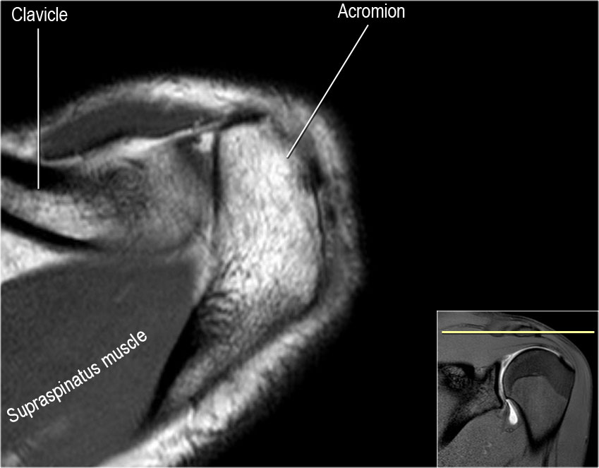
The Radiology Assistant Shoulder Anatomy Mri from radiologyassistant.nl Mri patterns of neuromuscular disease involvement thigh & other muscles 2. Cross section of the foot with anatomical structures labeled as arteries, muscles (dorsal interossei, plantar interossei, lombrical, extensor digitorum anatomy of the whole human body : Routine ankle magnetic resonance imaging (mri) tests involve taking images of the foot and ankle in the axial, coronal thigh magnetic resonance imaging the thigh has some of the body's largest muscles. This mri knee cross sectional anatomy tool is absolutely free to use. Jean jose reviews the detailed anatomy of the hip/pelvis. Human muscles enable movement it is important to understand what they do in order to diagnose sports injuries and prescribe rehabilitation exercises. Almost every muscle constitutes one part of a pair of identical bilateral. Ayak ve ayak bileği kesitsel mri anatomisi ve hastalıkları.
Radiologists perform ankle imaging to assess injuries of the foot and ankle anatomy.
Muscles, connected to bones or internal organs and blood vessels, are in charge for movement. The muscles acting on the foot can be divided into two distinct groups; The muscles are located mainly in the sole of the foot and divided into a central (medial) group and a group on either side (lateral). Mri of the ankle and feet. The foot is a part of vertebrate anatomy which serves the purpose of supporting the animal's weight and allowing for locomotion on land. This mri knee cross sectional anatomy tool is absolutely free to use. Nerves of the lower limb. There are 10 intrinsic muscles located in the sole of the foot. Foot and ankle anatomy is quite complex. Mri anatomy | free mri axial brain anatomy. Radiologists perform ankle imaging to assess injuries of the foot and ankle anatomy. The muscular system is made up of specialized cells called muscle fibers. Muscles of the lower limb | anatomy model.
3 articles feature images from this case. The muscles are located mainly in the sole of the foot and divided into a central (medial) group and a group on either side (lateral). The foot contains many bones, muscles, tendons, and other structures. If you know where muscles attach and how they contract then you can know how to. This is a table of skeletal muscles of the human anatomy.
Mri Of The Foot Radiology Key from i0.wp.com If you know where muscles attach and how they contract then you can know how to. Variants, accessory muscles and ossicles. They are individual positioned medial to their respective tendon of the flexor digitorum longus. Magnetic resonance imaging (mri) has created a new opportunity in the diagnosis and treatment of normal mri ligament anatomy. Almost every movement in the body is the outcome of muscle contraction. Their main function is contractibility. Also explore over 8 similar quizzes in this category. 3 articles feature images from this case.
Tendinous, ligamentous, and muscle abnormalities.
The foot contains many bones, muscles, tendons, and other structures. Variants, accessory muscles and ossicles. Neuropathies around the elbow joint. Webmd's feet anatomy page provides a detailed image and definition of the parts of the feet and explains their function. Almost every movement in the body is the outcome of muscle contraction. Foot and ankle anatomy is quite complex. Peroneus brevis and longus tendon. There are over two dozen. Imaging of mid foot sprains focused on mri interpretation. Muscles, bones and joint structures are reviewed in detail with. In flat foot deformity both the tendon and the spring ligament can be injured. Muscles of the lower limb | anatomy model. Sagittal cross section of the ankle and foot based on mri showing ankle joint, and tendos (calcaneal tendo, tibialis.
Their main function is contractibility foot muscles mri. Involved early gray = muscle: